Electromyography (EMG) is a diagnostic procedure used to assess the health and function of muscles and the nerve cells that control them, known as motor neurons. By measuring the electrical activity produced by skeletal muscles, EMG helps detect neuromuscular abnormalities, aiding in the diagnosis of various conditions affecting muscle and nerve function. This test is crucial for identifying disorders such as muscular dystrophy, peripheral neuropathies, and motor neuron diseases, enabling timely and targeted treatment.
To understand EMG, it is essential to grasp the basic anatomy and physiology of the neuromuscular system. Muscles contract in response to electrical signals transmitted by motor neurons. These neurons originate in the spinal cord and brain and extend their axons to muscle fibers. When a motor neuron fires, it generates an electrical impulse that causes the muscle fibers it innervates to contract.
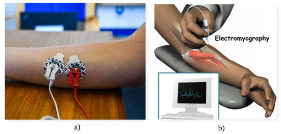
EMG records this electrical activity either at rest or during muscle contraction. The two primary types of electrodes used are:
The electrical signals captured provide insight into the muscle’s ability to respond to nerve stimulation and the integrity of the nerve pathways.
EMG is not a disease but a diagnostic tool used to investigate various neuromuscular disorders. Common causes or conditions for which EMG is indicated include:
Risk factors for these conditions vary but often include chronic diseases (e.g., diabetes), trauma, infections, autoimmune disorders, and genetic predispositions.
Patients undergoing EMG typically present with symptoms suggestive of neuromuscular dysfunction, including:
These symptoms help clinicians decide when EMG testing is appropriate.
EMG is part of a comprehensive diagnostic workup that includes:
The combination of EMG and NCS helps differentiate between muscle disorders, nerve disorders, and problems at the neuromuscular junction.
EMG itself is a diagnostic test and does not treat conditions. However, the results guide treatment decisions, which may include:
The choice of treatment depends on the underlying diagnosis revealed by EMG and other investigations.
The EMG procedure typically involves the following steps:
The procedure usually takes 30 to 60 minutes and may cause mild discomfort.
Since EMG is a diagnostic test rather than a surgical procedure, postoperative care is minimal. Patients may experience slight soreness or bruising at the needle insertion sites, which typically resolves within a day or two. Recommendations include:
Rehabilitation and recovery depend on the underlying condition diagnosed through EMG.
EMG is generally safe, but potential risks include:
Proper technique and sterile conditions minimize these risks.
The prognosis depends on the underlying neuromuscular condition diagnosed by EMG. Early and accurate diagnosis often leads to better management and improved outcomes. For example:
EMG plays a vital role in guiding prognosis by clarifying the nature and extent of neuromuscular involvement.
Seek medical evaluation and possible EMG testing if you experience:
Early consultation helps identify treatable conditions and prevent complications.
Electromyography (EMG) is a valuable diagnostic tool that provides critical information about muscle and nerve function. By detecting electrical abnormalities in muscles, EMG helps diagnose a wide range of neuromuscular disorders, guiding effective treatment strategies. While the procedure is generally safe and well-tolerated, it requires skilled interpretation by healthcare professionals. If you experience symptoms suggestive of neuromuscular problems, consult a healthcare provider to determine whether EMG testing is appropriate. Early diagnosis and intervention can significantly improve quality of life and functional outcomes.
Aenean porta orci nam commodo felis hac ridiculus fusce fames maximus erat sed dictumst blandit arcu suspendisse sollicitudin luctus in nec