Kamala Devi Hospital

Tumor Excision from Bone or Soft Tissue

Tumor Excision from Bone or Soft Tissue

Introduction

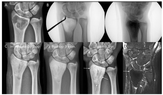
Tumors arising in bone or soft tissue can be benign or malignant and present unique challenges in diagnosis and management. Surgical excision remains a cornerstone in the treatment of many such tumors, aiming to remove the lesion completely while preserving as much function as possible. The approach to tumor excision depends on the tumor type, location, size, involvement of surrounding structures, and the patient’s overall health.

This article provides an overview of tumor excision from bone and soft tissue, including indications, preoperative evaluation, surgical techniques, and postoperative care.

Types of Tumors in Bone and Soft Tissue

Bone Tumors

  • Benign: Osteochondroma, enchondroma, giant cell tumor, osteoid osteoma.
  • Malignant: Osteosarcoma, Ewing’s sarcoma, chondrosarcoma, metastatic lesions.

Soft Tissue Tumors

  • Benign: Lipoma, hemangioma, neurofibroma, fibroma.
  • Malignant: Soft tissue sarcomas such as liposarcoma, synovial sarcoma, malignant peripheral nerve sheath tumor.

The biological behavior of the tumor dictates the urgency and extent of surgical excision.

Indications for Tumor Excision

  • Diagnostic biopsy: To obtain tissue for histopathological diagnosis.
  • Curative intent: Complete removal of benign or localized malignant tumors.
  • Palliative: To relieve pain, prevent pathological fractures, or improve function in advanced disease.
  • Prevention of complications: Such as neurovascular compression or infection.

Preoperative Assessment and Planning

Clinical Evaluation

  • Detailed history including duration, growth rate, pain, and systemic symptoms.
  • Physical examination focusing on size, consistency, mobility, neurovascular status, and involvement of adjacent structures.

Imaging Studies

  • X-rays: Initial assessment of bone involvement and lesion characteristics.
  • MRI: Gold standard for soft tissue delineation and extent of tumor.
  • CT scan: Useful for cortical bone involvement and surgical planning.
  • Bone scan or PET scan: To assess for metastasis or multifocal disease.

Biopsy

  • Essential for diagnosis before definitive surgery.
  • Can be performed as fine needle aspiration, core needle biopsy, or open biopsy.
  • Should be planned carefully to avoid contamination of uninvolved tissues.

Multidisciplinary Approach

  • Collaboration with oncologists, radiologists, pathologists, and rehabilitation specialists.
  • Preoperative chemotherapy or radiotherapy may be indicated for certain malignant tumors.

Surgical Techniques for Tumor Excision

The goal of surgery is complete tumor removal with negative margins while preserving function.

1. Excision of Bone Tumors

  • Intralesional Curettage: Removal of tumor tissue from within the bone cavity, often used for benign tumors like giant cell tumor.
  • Marginal Excision: Removal of the tumor with a margin of normal tissue, suitable for some benign or low-grade malignant tumors.
  • Wide Excision: Removal of the tumor with a cuff of normal tissue, standard for malignant tumors.
  • Radical Excision: Removal of the entire bone or compartment, reserved for extensive malignancies.

Reconstruction may be necessary after excision, using bone grafts, endoprostheses, or allografts.

2. Excision of Soft Tissue Tumors

  • Marginal Excision: Removal of the tumor along its pseudocapsule, often for benign lesions.
  • Wide Excision: Removal of the tumor with a margin of healthy tissue, required for malignant tumors.
  • Compartmental Resection: Removal of the entire anatomical compartment containing the tumor, used in aggressive malignancies.

Preservation of neurovascular structures is critical; sometimes sacrifice is necessary for oncological clearance.

3. Minimally Invasive and Advanced Techniques

  • Use of intraoperative imaging and navigation.
  • Limb-sparing surgeries to avoid amputation.
  • Use of tourniquets and meticulous hemostasis to reduce blood loss.

Intraoperative Considerations

  • Proper patient positioning and surgical exposure.
  • Identification and protection of vital structures.
  • Use of frozen section pathology to confirm margins.
  • Hemostasis and prevention of tumor seeding.

Postoperative Care and Rehabilitation

  • Pain management and wound care.
  • Early mobilization and physical therapy to restore function.
  • Monitoring for complications such as infection, hematoma, or neurovascular injury.
  • Regular follow-up with imaging to detect recurrence.

Complications

  • Local recurrence due to incomplete excision.
  • Infection, especially in large resections or reconstructions.
  • Neurovascular injury leading to functional deficits.
  • Pathological fractures in bone tumor excisions.
  • Lymphedema or wound healing problems in soft tissue tumor surgery.

Prognosis

  • Dependent on tumor type, grade, size, and completeness of excision.
  • Benign tumors generally have excellent outcomes.
  • Malignant tumors require multimodal treatment and have variable prognosis.
  • Early detection and appropriate surgical management improve survival and quality of life.

Conclusion

Tumor excision from bone or soft tissue is a complex surgical endeavor requiring careful planning and execution. Advances in imaging, surgical techniques, and multidisciplinary care have significantly improved outcomes. The primary objective remains complete tumor removal with preservation of function, minimizing morbidity, and preventing recurrence. Ongoing research and innovation continue to enhance the management of these challenging conditions.

Welcome to KamalaDevi Hospital

Aenean porta orci nam commodo felis hac ridiculus fusce fames maximus erat sed dictumst blandit arcu suspendisse sollicitudin luctus in nec

Make an Appointment.