Magnetic Resonance Imaging (MRI) is a non-invasive medical imaging technique widely used to visualize detailed internal structures of the body. Unlike X-rays or CT scans, MRI uses strong magnetic fields and radio waves to generate high-resolution images of organs, tissues, and bones without exposure to ionizing radiation. This technology plays a crucial role in diagnosing a broad range of conditions, from neurological disorders to musculoskeletal injuries and cancers. Its ability to provide clear contrast between different soft tissues makes MRI indispensable in modern medicine.
MRI capitalizes on the magnetic properties of hydrogen atoms, which are abundant in the human body due to water and fat content. When placed in a strong magnetic field, these hydrogen nuclei align with the field. Radiofrequency pulses then disturb this alignment, and as the nuclei return to their original state, they emit signals captured by the MRI scanner. These signals are processed to create detailed images.
Understanding basic anatomy is essential to appreciate MRI’s utility. For example, in brain imaging, MRI can differentiate gray matter, white matter, cerebrospinal fluid, and pathological lesions. In musculoskeletal imaging, it can distinguish muscles, ligaments, cartilage, and bone marrow. This contrast resolution is vital for accurate diagnosis.
MRI is a key diagnostic tool used after clinical examination and initial investigations. The diagnostic process includes:
MRI may be combined with contrast agents (e.g., gadolinium) to enhance visualization of blood vessels or tumors.
MRI itself is not a treatment but informs treatment decisions. Based on MRI findings, treatment options may include:
MRI is also used intraoperatively or for image-guided interventions in some cases.
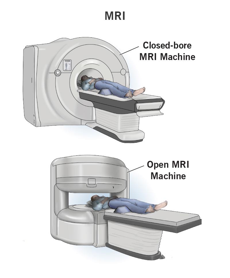
The MRI procedure involves several steps:
Since MRI is a diagnostic procedure, postoperative care is not applicable. However, if MRI guides surgery or intervention, postoperative care depends on the specific treatment performed. Rehabilitation may include physical therapy, medication, and follow-up imaging to monitor recovery.
MRI is generally safe but has some risks and limitations:
No ionizing radiation is involved, making MRI safer than CT in many cases.
MRI improves prognosis by enabling early and accurate diagnosis, which facilitates timely and appropriate treatment. It helps monitor disease progression and response to therapy, contributing to better patient outcomes.
Magnetic Resonance Imaging is a powerful diagnostic tool that provides detailed images of the body’s internal structures without radiation exposure. It plays a vital role in diagnosing a wide range of medical conditions, guiding treatment decisions, and monitoring outcomes. While generally safe, MRI requires careful patient screening and preparation. If you experience symptoms that warrant detailed internal imaging, consult a healthcare professional to discuss whether MRI is the right choice for you. Early diagnosis through MRI can significantly improve treatment success and quality of life.
Aenean porta orci nam commodo felis hac ridiculus fusce fames maximus erat sed dictumst blandit arcu suspendisse sollicitudin luctus in nec