A DEXA scan, short for Dual-Energy X-ray Absorptiometry, is a specialized imaging test primarily used to measure bone mineral density (BMD). It is considered the gold standard for diagnosing osteoporosis and assessing fracture risk. Given the increasing prevalence of osteoporosis and related fractures worldwide, the DEXA scan plays a crucial role in early detection, prevention, and management of bone health issues. This non-invasive, quick, and low-radiation procedure helps healthcare providers make informed decisions about treatment and lifestyle modifications to maintain bone strength and reduce the risk of debilitating fractures.
To understand the importance of a DEXA scan, it is helpful to know some basic bone anatomy and physiology. Bones are living tissues composed mainly of a matrix of collagen fibers and minerals, primarily calcium and phosphate, which provide strength and rigidity. Bone remodeling is a continuous process where old bone is resorbed by cells called osteoclasts and new bone is formed by osteoblasts. This balance maintains bone density and structural integrity.
Diagnosis of low bone density or osteoporosis involves a combination of clinical assessment and imaging:
The results are expressed as:
Additional tests may include blood tests to rule out secondary causes of bone loss.
Treatment aims to prevent fractures by improving bone density and reducing risk factors:
Surgery is generally reserved for fracture management rather than osteoporosis itself. Procedures may include vertebroplasty or kyphoplasty for vertebral fractures or orthopedic fixation for hip and wrist fractures.
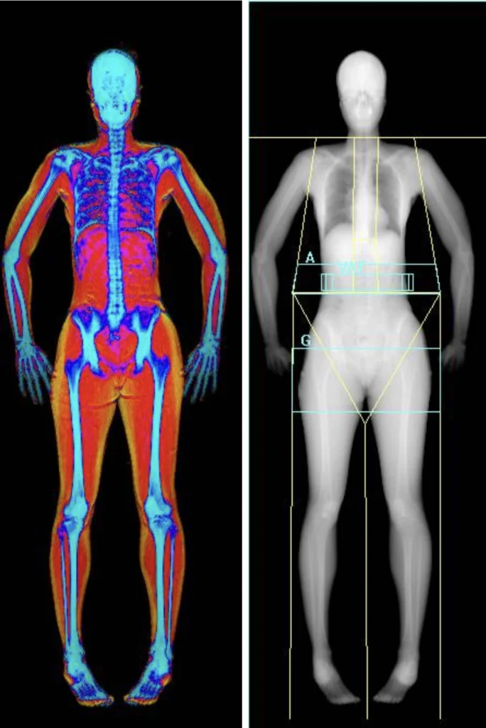
The DEXA scan procedure is straightforward and painless:
The radiation exposure is minimal, much less than a standard chest X-ray.
Since the DEXA scan is non-invasive, no postoperative care is required. However, if the scan leads to diagnosis of osteoporosis or fractures, follow-up care may include:
The DEXA scan is very safe with minimal risks:
No surgical or invasive risks are associated with the scan itself.
Early detection of low bone density through DEXA scanning allows timely intervention, significantly reducing the risk of fractures and associated complications. With appropriate treatment and lifestyle changes, many patients maintain good bone health and quality of life. Untreated osteoporosis, however, can lead to debilitating fractures, chronic pain, and loss of independence.
The DEXA scan is a vital diagnostic tool in assessing bone mineral density and managing osteoporosis. It provides accurate, quick, and safe measurement of bone health, enabling early detection and intervention. Understanding risk factors, symptoms, and treatment options empowers patients to take proactive steps toward maintaining strong bones. If you or a loved one are at risk for osteoporosis, consult a healthcare professional about the benefits of a DEXA scan and appropriate bone health strategies. Early action can preserve mobility, reduce fracture risk, and improve overall quality of life.
Aenean porta orci nam commodo felis hac ridiculus fusce fames maximus erat sed dictumst blandit arcu suspendisse sollicitudin luctus in nec