Kamala Devi Hospital

Fluoroscopy: A Comprehensive Overview

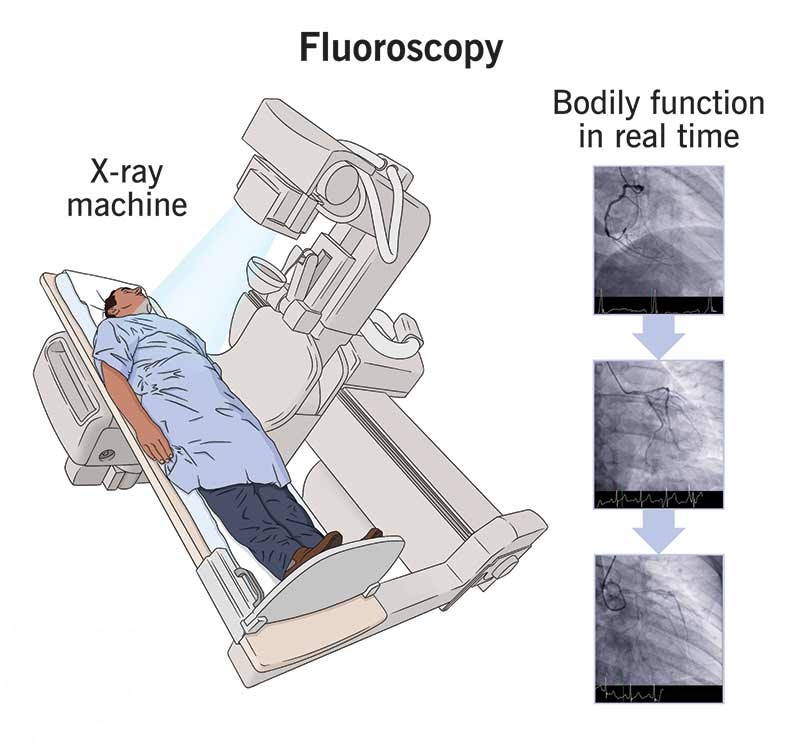
Fluoroscopy is a medical imaging technique that provides real-time moving images of the internal structures of the body using X-rays. Unlike static X-ray images, fluoroscopy allows physicians to observe dynamic processes such as the movement of joints, the flow of contrast agents through blood vessels, or the function of the gastrointestinal tract. This capability makes fluoroscopy an invaluable tool in diagnosis, treatment planning, and guiding minimally invasive procedures.

The importance of fluoroscopy lies in its ability to offer continuous visualization, which enhances the accuracy of interventions and improves patient outcomes. It is widely used in various medical specialties including orthopedics, cardiology, gastroenterology, and interventional radiology.

Anatomy / Background

Understanding fluoroscopy requires a basic knowledge of the anatomical regions commonly examined with this technique. For example:

  • Musculoskeletal system: Fluoroscopy can visualize joint movement, bone alignment, and guide fracture reductions.
  • Cardiovascular system: It helps in visualizing blood flow through arteries and veins during angiography.
  • Gastrointestinal tract: Fluoroscopy tracks the passage of contrast agents through the esophagus, stomach, intestines, and colon to assess motility and detect abnormalities.
  • Urinary system: It is used in procedures like cystography to evaluate bladder function.
Signup our newsletter to get update information, news, insight or promotions.

Causes / Etiology

Symptoms / Clinical Presentation

Diagnosis

The diagnostic process involving fluoroscopy includes:

  1. Clinical Examination: A thorough history and physical examination to identify symptoms and signs.
  2. Imaging Studies: Fluoroscopy is performed using specialized equipment in a radiology suite. The patient may be asked to swallow, move, or hold certain positions depending on the study.
  3. Use of Contrast Agents: To highlight specific organs or vessels.
  4. Additional Tests: Such as blood work or other imaging modalities (MRI, CT) may complement fluoroscopic findings.

The real-time imaging allows clinicians to observe physiological processes and make accurate diagnoses.

Treatment Options

Fluoroscopy is not a treatment but a diagnostic and procedural aid. However, it plays a crucial role in guiding treatments such as:

  • Non-surgical treatments: Including guided injections (e.g., corticosteroids into joints or spine), catheter placements, and biopsies.
  • Surgical treatments: Fluoroscopy assists in minimally invasive surgeries like angioplasty, stent placement, orthopedic fixation, and gastrointestinal interventions.

The choice of treatment depends on the diagnosis and patient-specific factors.

Procedure Details

When fluoroscopy is used for procedural guidance, the steps typically include:

  1. Preparation: The patient is positioned on the fluoroscopy table. Consent and explanation of the procedure are obtained.
  2. Sterilization and Local Anesthesia: If an invasive procedure is planned.
  3. Contrast Administration: Oral, intravenous, or intra-arterial contrast may be given.
  4. Real-time Imaging: The physician monitors the fluoroscopic images to guide instruments or assess function.
  5. Completion: Once the procedure is done, the equipment is removed, and the patient is monitored.

The procedure duration varies depending on the complexity.

Postoperative Care / Rehabilitation

Post-procedure care depends on the intervention performed:

  • Observation: Monitoring for immediate complications such as bleeding or allergic reactions to contrast.
  • Pain Management: As needed.
  • Activity Restrictions: Based on the procedure.
  • Follow-up Imaging: To assess treatment success.
  • Rehabilitation: Physical therapy may be recommended for musculoskeletal interventions.

Patients are advised to report any unusual symptoms promptly.

Risks and Complications

While fluoroscopy is generally safe, potential risks include:

  • Radiation Exposure: Prolonged fluoroscopy increases radiation dose; protective measures are essential.
  • Allergic Reactions: To contrast agents.
  • Infection or Bleeding: If invasive procedures are performed.
  • Tissue Damage: Rarely, due to radiation or instrument manipulation.

Healthcare providers take precautions to minimize these risks.

Prognosis

The prognosis depends on the underlying condition diagnosed or treated with fluoroscopy. Early and accurate diagnosis facilitated by fluoroscopy often leads to better outcomes. Minimally invasive procedures guided by fluoroscopy typically result in shorter recovery times and fewer complications compared to open surgery.

When to See a Doctor

conclusion

Fluoroscopy is a vital imaging modality that provides dynamic visualization of internal body structures, aiding in diagnosis and guiding a variety of medical procedures. Its ability to offer real-time images enhances the precision of interventions and improves patient care. Understanding the applications, procedure details, and associated risks of fluoroscopy empowers patients and healthcare providers to make informed decisions. If you or a loved one requires fluoroscopic evaluation or treatment, consult with a qualified healthcare professional to ensure safe and effective care.

Explore More:

Welcome to KamalaDevi Hospital

Aenean porta orci nam commodo felis hac ridiculus fusce fames maximus erat sed dictumst blandit arcu suspendisse sollicitudin luctus in nec

Make an Appointment.