Kamala Devi Hospital

Growth Plate Fracture Fixation

Growth Plate Fracture Fixation: Understanding Treatment and Recovery

Introduction

Growth plate fractures, also known as physeal fractures, are injuries that occur in the growth plates of children and adolescents. The growth plate (physis) is a layer of cartilage near the ends of long bones where bone growth occurs. Because these plates are weaker than the surrounding bone and ligaments, they are susceptible to fractures during trauma.

Proper management of growth plate fractures is critical to ensure normal bone growth and to prevent complications such as growth arrest, deformity, or limb length discrepancy. Growth plate fracture fixation is a surgical procedure used to stabilize displaced or unstable fractures of the growth plate, promoting healing and preserving growth potential.

This article provides an in-depth overview of growth plate fractures, indications for fixation, surgical techniques, postoperative care, and outcomes.

Understanding Growth Plate Fractures

Anatomy of the Growth Plate

The growth plate is composed of cartilage cells that proliferate and ossify to allow longitudinal bone growth. It is located between the metaphysis (shaft end) and the epiphysis (bone end). Because it is the last portion of the bone to ossify, it remains vulnerable to injury during childhood and adolescence.

Clubfoot happens because of an issue with your child’s tendons, the tissues that connect muscle to bone. The tendons in your baby’s leg and foot are shorter and tighter than they should be. That causes the foot to twist. About half of babies with clubfoot have an issue with both feet.

Types of Growth Plate Fractures

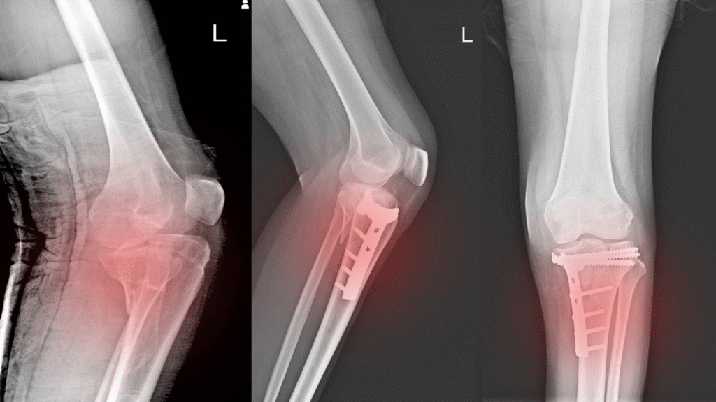
Growth plate fractures are classified by the Salter-Harris system, which helps guide treatment and prognosis:

  • Type I: Fracture through the growth plate without involving bone.
  • Type II: Fracture through the growth plate and metaphysis.
  • Type III: Fracture through the growth plate and epiphysis.
  • Type IV: Fracture through the metaphysis, growth plate, and epiphysis.
  • Type V: Compression injury to the growth plate.

Types III and IV often require surgical fixation due to involvement of the joint surface and risk of growth disturbance.

Indications for Growth Plate Fracture Fixation

Not all growth plate fractures require surgery. Indications for fixation include:

  • Displaced fractures: Where bone fragments are misaligned.
  • Intra-articular fractures: Involving the joint surface (Salter-Harris III and IV).
  • Unstable fractures: At risk of displacement with conservative treatment.
  • Open fractures: Where the skin is broken.
  • Failure of closed reduction: When non-surgical realignment is unsuccessful.
  • Fractures with neurovascular compromise.

The goal of fixation is to restore anatomical alignment, stabilize the fracture, and minimize damage to the growth plate.

Preoperative Evaluation

  • Clinical assessment: Includes history of injury, physical examination for swelling, deformity, and neurovascular status.
  • Imaging: X-rays are essential to classify the fracture and plan treatment. CT or MRI may be used for complex fractures.
  • Planning: Determining the surgical approach and fixation method based on fracture type and location

Surgical Techniques for Growth Plate Fracture Fixation

Anesthesia and Positioning

Surgery is performed under general anesthesia. The patient is positioned to allow optimal access to the fracture site.

Reduction

  • Closed reduction: Attempted first by manipulating the bone fragments without incision.
  • Open reduction: Required if closed reduction fails or if the fracture is complex.

Fixation Methods

The choice of fixation depends on the fracture type, size, and location:

  • Smooth Kirschner wires (K-wires): Commonly used for temporary fixation; they avoid crossing the growth plate or minimize damage.
  • Screws: Used cautiously; if crossing the growth plate, screws should be placed perpendicular to minimize growth disturbance.
  • Bioabsorbable pins: Sometimes used to avoid hardware removal.
  • Plates: Rarely used in growth plate fractures due to risk of growth interference.

Surgical Steps

  1. Incision and exposure: If open reduction is needed.
  2. Anatomic reduction: Precise realignment of the fracture fragments.
  3. Fixation: Stabilizing the fracture with appropriate hardware.
  4. Verification: Intraoperative imaging to confirm alignment and hardware placement.
  5. Closure: Layered closure of soft tissues.

Postoperative Care

  • Immobilization: Typically with a cast or splint to protect the fixation and allow healing.
  • Weight-bearing: Restricted initially; gradual progression based on healing.
  • Pain management: Using medications as needed.
  • Follow-up: Regular clinical and radiographic evaluations to monitor healing and detect complications.

Complications and Management

Growth plate fractures carry a risk of complications, especially if not properly treated:

  • Growth arrest: Premature closure of the growth plate leading to limb length discrepancy or angular deformity.
  • Malunion or nonunion: Improper healing causing deformity or instability.
  • Joint stiffness: Particularly in intra-articular fractures.
  • Infection: Postoperative wound infection.
  • Neurovascular injury: Rare but serious.
  • Early detection and management of complications are essential to optimize outcomes.

Outcomes and Prognosis

The prognosis of growth plate fractures depends on:

  • Fracture type and severity.
  • Accuracy of reduction and fixation.
  • Patient age and remaining growth potential.
  • Promptness of treatment.

With appropriate fixation and care, most children regain full function with minimal long-term issues. However, close monitoring until skeletal maturity is important to identify and address any growth disturbances.

Conclusion

Growth plate fracture fixation is a critical intervention for displaced or unstable physeal fractures in children and adolescents. By restoring anatomical alignment and stabilizing the fracture, this procedure helps preserve the growth potential of the bone and prevents deformities.

Early diagnosis, careful surgical technique, and diligent postoperative follow-up are key to successful outcomes. Parents and caregivers should seek immediate medical attention for suspected growth plate injuries to ensure timely and effective treatment.

Welcome to KamalaDevi Hospital

Aenean porta orci nam commodo felis hac ridiculus fusce fames maximus erat sed dictumst blandit arcu suspendisse sollicitudin luctus in nec

Make an Appointment.