Kamala Devi Hospital

Microdiscectomy: A Comprehensive Overview

Microdiscectomy is a minimally invasive surgical procedure designed to relieve pressure on spinal nerves caused by herniated discs, primarily in the lumbar (lower back) region. It is one of the most common surgeries performed to treat sciatica and other nerve compression symptoms resulting from disc herniation. This procedure is important because it offers significant pain relief, improved mobility, and a quicker recovery compared to traditional open spine surgeries. Understanding microdiscectomy helps patients make informed decisions about their treatment options for back and leg pain related to disc problems.

Anatomy / Background

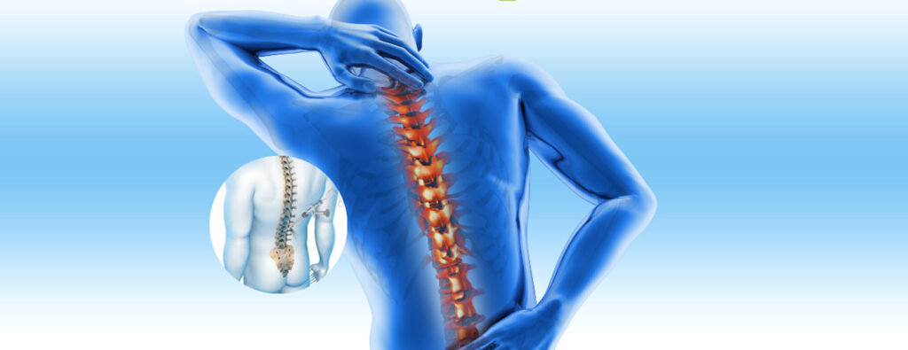
The spine is composed of vertebrae stacked on top of each other, separated by intervertebral discs that act as shock absorbers. Each disc has a tough outer layer called the annulus fibrosus and a gel-like center called the nucleus pulposus. Nerves exit the spinal cord through spaces between vertebrae called foramina. When a disc herniates, the nucleus pulposus protrudes through a tear in the annulus fibrosus, potentially compressing nearby spinal nerves.

In the lumbar spine, nerve compression often causes pain, numbness, or weakness radiating down the leg, a condition known as sciatica. Microdiscectomy targets the herniated portion of the disc to relieve this nerve pressure.

Causes / Etiology

Symptoms / Clinical Presentation

Patients with a herniated lumbar disc typically present with:

  • Lower Back Pain: Often localized but can be mild compared to leg symptoms.
  • Radicular Pain (Sciatica): Sharp, shooting pain radiating from the lower back down one leg, following the path of the compressed nerve.
  • Numbness or Tingling: Sensory changes in the leg or foot.
  • Muscle Weakness: Difficulty lifting the foot or toes (foot drop) or weakness in leg muscles.
  • Reflex Changes: Decreased reflexes in the knee or ankle.
  • Worsening Symptoms with Movement: Pain may increase with sitting, bending, coughing, or sneezing.

Diagnosis

Diagnosis of a herniated disc requiring microdiscectomy involves:

  • Clinical Examination: Assessment of pain distribution, neurological deficits (strength, sensation, reflexes), and special tests such as the straight leg raise test.
  • Imaging Studies:
    • Magnetic Resonance Imaging (MRI): The gold standard to visualize disc herniation and nerve compression.
    • Computed Tomography (CT) Scan: Used if MRI is contraindicated.
    • X-rays: To rule out other causes like fractures or instability.
  • Electrodiagnostic Tests: Nerve conduction studies or electromyography (EMG) may be used to assess nerve function.

Treatment Options

Non-Surgical Treatments

Most patients initially try conservative management, including:

  • Physical Therapy: Exercises to strengthen back muscles and improve flexibility.
  • Medications: Nonsteroidal anti-inflammatory drugs (NSAIDs), muscle relaxants, or oral steroids.
  • Epidural Steroid Injections: To reduce inflammation around the nerve root.
  • Activity Modification: Avoiding aggravating activities and using ergonomic supports.
Surgical Treatment: Microdiscectomy

Surgery is considered when:

  • Symptoms persist beyond 6 weeks despite conservative care.
  • Severe or progressive neurological deficits occur.
  • Intractable pain significantly impairs quality of life.

Procedure Details

Microdiscectomy is performed under general anesthesia and involves the following steps:

  1. Patient Positioning: The patient lies face down on the operating table.
  2. Incision: A small (approximately 1-2 inch) incision is made over the affected vertebral level.
  3. Muscle Retraction: Paraspinal muscles are gently retracted to expose the vertebrae.
  4. Microscopic Visualization: A surgical microscope or magnifying loupes are used for precision.
  5. Laminotomy: A small portion of the lamina (bony arch) is removed to access the spinal canal.
  6. Removal of Herniated Disc Material: The surgeon carefully removes the herniated nucleus pulposus compressing the nerve root.
  7. Nerve Root Decompression: Ensuring the nerve is free from pressure.
  8. Closure: The incision is closed with sutures or staples.

The minimally invasive nature of microdiscectomy allows for less tissue damage, reduced blood loss, and faster recovery compared to traditional open discectomy.

Postoperative Care / Rehabilitation

  • Hospital Stay: Usually outpatient or 1-day admission.
  • Pain Management: Oral analgesics and anti-inflammatory medications.
  • Activity: Early mobilization is encouraged; avoid heavy lifting and twisting for several weeks.
  • Physical Therapy: Begins within a few weeks to restore strength and flexibility.
  • Follow-Up: Regular visits to monitor wound healing and neurological status.
  • Return to Work: Typically within 4-6 weeks, depending on job demands.

Risks and Complications

Though generally safe, microdiscectomy carries potential risks:

  • Infection: At the surgical site or deeper tissues.
  • Bleeding or Hematoma Formation
  • Nerve Injury: Leading to persistent pain, numbness, or weakness.
  • Dural Tear: Leakage of cerebrospinal fluid.
  • Recurrence of Disc Herniation: At the same or adjacent level.
  • Persistent or Incomplete Symptom Relief
  • Anesthesia-Related Complications

Prognosis

The prognosis after microdiscectomy is generally excellent:

  • Approximately 80-90% of patients experience significant pain relief.
  • Most regain normal function and return to daily activities.
  • Recurrence rates are low but possible.
  • Early surgery in appropriate candidates often leads to better outcomes.

When to See a Doctor

Conclusion

Microdiscectomy is a highly effective surgical option for patients suffering from nerve compression due to herniated lumbar discs. Understanding the anatomy, causes, symptoms, and treatment options empowers patients to make informed decisions. While many cases improve with non-surgical care, microdiscectomy offers rapid relief and functional recovery when surgery is indicated. If you experience persistent back and leg pain or neurological symptoms, consult a healthcare professional for proper evaluation and personalized treatment planning. Early diagnosis and appropriate intervention can significantly improve quality of life and long-term outcomes.

Welcome to KamalaDevi Hospital

Aenean porta orci nam commodo felis hac ridiculus fusce fames maximus erat sed dictumst blandit arcu suspendisse sollicitudin luctus in nec

Make an Appointment.